
A Patient-Advocate's Guide to Recognizing Thoracic endometriosis
If you've ever doubled over because of your period with sharp chest & shoulder pain that felt impossible to explain, or coughed up blood and were told it was "just a chest infection," or had a sudden collapsed lung in your 20s or 30s and no one could figure out why — this post is for you
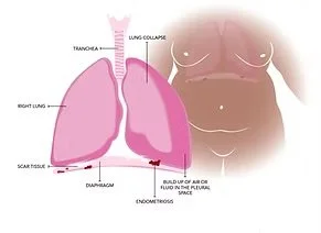
Thoracic Endometriosis Syndrome (TES) is the most common manifestation of endometriosis outside the pelvis. It involves endometrial-like tissue growing on or around the diaphragm, lungs, airways, and the lining of the chest. Despite affecting an estimated 80% of patients that also have severe pelvic endometriosis (Stages III & IV), TES remains one of the most underdiagnosed, misdiagnosed, and dismissed conditions in modern medicine, frequently delayed by an average of 19 months from a patient's first thoracic event to a correct diagnosis, often years longer when factored alongside the standard 7-to-11-year endometriosis diagnostic delay. This post is based on our recently completed chapter, "Thoracic Endometriosis Syndrome" (Amirlatifi, Niehaus, Najmi, Ambarge, Sirjani, and Nezhat), a comprehensive review of the current science, clinical presentation, and surgical management of TES. We are translating that academic work into plain language here, so that patients, families, and advocates have the tools to recognize TES, demand appropriate evaluation, and stop being dismissed.
WHAT IS THORACIC ENDOMETRIOSIS SYNDROME?
Endometriosis is a chronic, systemic, estrogen-driven, inflammatory disease defined by endometrial-like glands and stroma growing outside the uterus. While it predominantly affects the pelvic cavity, the thorax: the chest cavity containing the lungs, diaphragm, and surrounding pleura, is the most common site for endometriosis to appear beyond the pelvis. Thoracic Endometriosis Syndrome encompasses a wide spectrum of presentations, including:
— Catamenial pneumothorax (collapsed lung during menstruation)
— Catamenial hemothorax (blood in the chest cavity during menstruation)
— Catamenial hemoptysis (coughing up blood during menstruation)
— Pulmonary nodules
— Diaphragmatic endometriosis (often presenting as shoulder, neck, or upper abdominal pain)
— Diaphragmatic hernia
— Recurrent pleural effusion
The word "catamenial" simply means "occurring in relation to menstruation" — typically within 24 hours before or 72 hours after a period begins. However, more recent research has shown that thoracic endometriosis can also occur outside of this menstrual window, which has prompted experts to propose the broader term thoracic endometriosis-related pneumothorax (TERP) to capture cases that don't fit the textbook timing.
A BRIEF HISTORY: 1938 TO TODAY
The earliest reports of thoracic endometriosis date back to 1938, when Schwarz first documented pulmonary parenchymal disease. Barnes described endometriosis-related hemothorax in 1953, and Maurer reported on diaphragmatic endometriosis surgery in 1958. Building on these early open-surgical reports, Dr. Camran Nezhat pioneered the minimally invasive surgical management of thoracic endometriosis in 1992, performing the world's first laparoscopic diaphragm resection and repair. That same year, he extended this approach to thoracic and pulmonary endometriosis, establishing minimally invasive surgeryas a viable surgical platform for endometriosis disease beyond the peritoneal cavity. This work fundamentally transformed what was once considered an open-chest, highly invasive procedure into something far safer, less traumatic, and more accessible for patients.
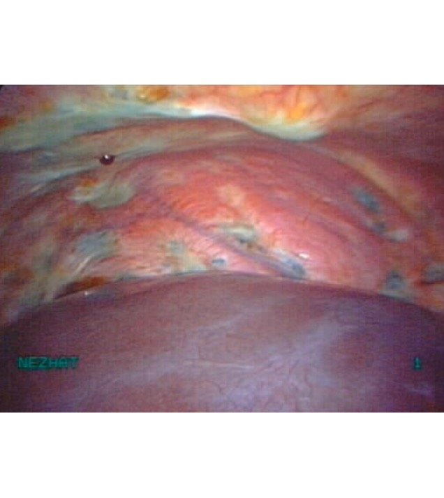
Nezhat Surgery
HOW COMMON IS TES, REALLY?
Up to 80% of patients with TES also have concurrent pelvic endometriosis — meaning the disease is rarely "thoracic only." Thoracic symptoms typically lag behind pelvic symptoms by 5 to 7 years, placing the mean age at TES diagnosis around 35. Among patients referred for surgical evaluation of spontaneous pneumothorax, up to 30% are ultimately found to have underlying thoracic endometriosis. That number alone should be enough to galvanize medical education reform — yet most pulmonologists and emergency physicians still do not routinely consider endometriosis when a person of reproductive age presents with a collapsed lung. Up to 70% of TES patients remain entirely asymptomatic, which means the actual prevalence is likely much higher than reported. Diaphragmatic endometriosis specifically is reported in 0.19% to 1.5% of patients during pelvic surgery — but when surgeons perform systematic bilateral diaphragmatic inspection with liver mobilization, detection rates jump to 4.7% or higher. In other words: the disease is being missed because no one is looking.
THE THREE ANATOMICAL CATEGORIES OF TES
TES is typically grouped into three categories based on where the disease is found in the chest:
PLEURAL TES (THE LINING OF THE LUNG AND CHEST WALL)
Pleural thoracic endometriosis affects the visceral and parietal pleura — the thin membranes lining the lungs and the inside of the chest cavity. It most commonly presents as catamenial pneumothorax (a collapsed lung during menstruation) or catamenial hemothorax (blood accumulating in the chest cavity).
Symptoms can include:
— Pleuritic chest pain (sharp pain with breathing)
— Sudden shoulder pain
— Cough
— Shortness of breath
Clinically, catamenial pneumothorax is often indistinguishable from a standard spontaneous pneumothorax — which is why so many patients have multiple recurrences before anyone connects the timing to their menstrual cycle. Catamenial hemothorax can mimic pulmonary embolism, leading to unnecessary anticoagulation and missed diagnoses.
BRONCHOPULMONARY AND PARENCHYMAL TES (LUNG TISSUE ITSELF)
This less common variant involves endometriosis within the lung tissue or airways. It may present as:
— Catamenial hemoptysis (coughing up blood during menstruation)
— Pulmonary nodules detected incidentally on imaging Catamenial hemoptysis tends to affect younger patients and is rarely life-threatening — but it is frequently misdiagnosed as tuberculosis, lung infection, or even cancer before the menstrual pattern is recognized.
Pulmonary nodules are typically found in older patients, located in the lung periphery, and are usually discovered by accident during imaging for an unrelated reason.
DIAPHRAGMATIC TES (THE MUSCLE BENEATH THE LUNGS)
This is by far the most common form of thoracic endometriosis, identified in nearly 79% of patients evaluated thoracoscopically. The diaphragm is the dome-shaped muscle that separates your chest from your abdomen, and endometriotic implants on its surface can cause:
— Cyclic right shoulder pain (the most classic red flag)
— Neck or upper back pain
— Right upper quadrant abdominal pain
— Epigastric (upper-middle abdominal) pain —> The pain pattern is due to phrenic nerve irritation
— a nerve that travels from the diaphragm up to the shoulder, which is why diaphragmatic endometriosis so often presents as referred shoulder pain that feels disconnected from where the actual disease lives.
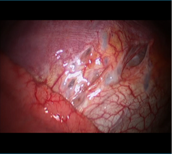
Over time, repeated cycles of inflammation and tissue breakdown on the diaphragm can produce small perforations that may merge into larger holes — eventually leading to diaphragmatic hernia, and in rare cases, full diaphragmatic rupture as the first sign of disease.
WHY DOES ENDO GO TO THE CHEST? THE LEADING THEORIES
There is no single explanation for how endometriosis reaches the chest. Rather, several mechanisms appear to be at work, often in combination:
1. Retrograde Menstruation with Transperitoneal Migration: Sampson's classic theory proposes that endometrial cells flow backward through the fallopian tubes into the peritoneal cavity, then travel along the right paracolic gutter — the natural rightward flow of peritoneal fluid — up to the diaphragm and through small congenital or acquired holes (fenestrations) into the chest. This explains the striking 9-to-1 right-sided predominance of thoracic disease.
2. Coelomic Metaplasia: First proposed by Meyer in 1924, this theory suggests that pleural and peritoneal mesothelial cells, which share an embryonic origin, can transform into endometrial-like tissue under hormonal and immune influence.
3. Lymphatic and Hematogenous Dissemination: Advanced by Halban in 1925 and supported by modern experimental models, this theory proposes that endometrial cells can travel through the lymph or bloodstream — which would explain the rare but documented cases of bilateral bronchopulmonary involvement.
4. Genetic and Epigenetic Factors: Genetic susceptibility accounts for an estimated 50% of disease risk. Recent molecular work has identified clonal expansion of endometrial stem cells carrying mutations in tumor suppressor genes such as ARID1A and PTEN, along with aberrant DNA methylation and histone remodeling. No single theory accounts for every case. The reality is likely a multifactorial cascade — which is why a one-size-fits-all approach to diagnosis and treatment continues to fail patients. HOW TES IS DIAGNOSED The diagnosis of TES is driven first and foremost by clinical suspicion. A thorough patient history is often more diagnostically powerful than any single test — yet patients are routinely sent home from emergency departments and pulmonology clinics without ever being asked whether their symptoms correlate with their menstrual cycle. Red flags every clinician should ask about:
— Symptom correlation with menses
— Recurrent right-sided thoracic events
— Reproductive age (typically 15–45) — Concurrent infertility
— Dysmenorrhea, dyspareunia, dyschezia, chronic pelvic pain
The freely available Endometriosis Risk Advisor app can serve as a useful non-invasive screening tool — particularly in primary care or emergency settings where access to a specialist may be delayed.
IMAGING
Chest X-ray or CT is typically the first imaging step in a suspected pneumothorax, but these have low specificity for endometriotic lesions and mostly serve to rule out other causes. MRI of the chest and abdomen, particularly with fat-suppressed T1-weighted sequences, is currently the most appropriate preoperative imaging modality. It achieves sensitivity of 78–83% for diaphragmatic endometriosis, with excellent interobserver agreement.
Dedicated thoracic MRI protocols with sagittal and coronal projections, plus breath-hold or respiratory-triggered techniques, significantly improve detection of small plaques. Scheduling MRI to coincide with menstruation may further enhance diagnostic yield, yet this timing is almost never done in routine clinical practice. The 2025 ESUR consensus guidelines introduced a standardized, compartment-based approach to reporting endometriosis on MRI — a small but meaningful step toward consistency. However, the panel did not reach agreement on whether systematic diaphragmatic evaluation should be incorporated into routine pelvic MRI protocols. We believe it should be.
BIOMARKERS CA-125
has been studied as a serum biomarker but has limited sensitivity (around 52%) and poor specificity, making it a weak standalone screening tool. That said, some authors advocate measuring CA-125 in any reproductive-age patient with recurrent pneumothorax, as an elevation may support the indication for diagnostic thoracoscopy. Newer biomarker research is promising. Circulating endometrial cells have been identified in the peripheral blood of patients with catamenial-suspicious pneumothorax. Serum microRNA panels have achieved diagnostic accuracies of 90% or higher in machine learning models.
A 2025 study identified differentially expressed microRNAs specific to adolescent endometriosis for the first time. Multiple diagnostic companies are developing blood-based, saliva-based, and menstrual-blood-based tests — none yet validated for routine clinical use, but a hopeful sign that less invasive diagnostic options are on the horizon.
SURGICAL DIAGNOSIS: VATS AND LAPAROSCOPY
Video-assisted thoracoscopic surgery (VATS) serves as both the diagnostic and therapeutic procedure of choice when imaging and biomarkers are inconclusive. It allows direct visualization and concurrent treatment. Critically, every video laparoscopy for pelvic endometriosis should include thorough diaphragmatic inspection.
"Sentinel lesions" — small superficial implants less than 1 cm on the right anterior diaphragmatic surface — should prompt examination of the entire diaphragm, as these are frequently the visible tip of much more extensive posterior disease hidden behind the right hepatic lobe. Liver mobilization is often required to actually see what's there.
HISTOLOGY AND IMMUNOHISTOCHEMISTRY
Histological diagnosis relies on identifying endometrial-like glands, stroma, and hemosiderin-laden macrophages on H&E staining — though in practice, thoracic specimens are often small, fibrotic, inflamed, or damaged by thermal artifact, making diagnosis difficult.
Immunohistochemistry — particularly with ER, PR, and CD10 antibodies — increases diagnostic yield from 61.7% to over 80%. Newer markers like IFITM1 (which demonstrated 100% positivity in extragenital endometriosis) and PAX8 are emerging as valuable adjuncts when CD10 is ambiguous.
As we've previously argued, every pleural, diaphragmatic, pulmonary, and bronchial specimen should undergo standard histological evaluation, with comprehensive immunohistochemistry routinely performed when clinical suspicion warrants. HOW TES IS TREATED Management of TES follows the same general principles as pelvic endometriosis: conservative medical therapy first, surgical intervention when medical treatment fails or is contraindicated.
MEDICAL MANAGEMENT
First-line options include:
— GnRH agonists
— Oral contraceptives (though with significant limitations, see below)
— Progestins (dienogest, norethindrone acetate)
— Aromatase inhibitors Danazol is no longer recommended as first-line therapy due to its significant androgenic side effects. Oral GnRH antagonists (relugolix, elagolix, linzagolix) have emerged as promising alternatives, with relugolix combination therapy showing sustained improvement in pelvic pain symptoms for up to 104 weeks in the SPIRIT extension study
— though data specific to TES remain limited. A critical note on oral contraceptives alone: Ciriaco et al. demonstrated a 100% catamenial pneumothorax recurrence rate when combined estrogen-progesterone oral contraceptives were used as the sole post-surgical treatment.
This is a finding that every clinician treating TES patients needs to internalize.
SURGICAL MANAGEMENT
When medical therapy fails, surgical excision is required. Our team's approach has evolved meaningfully over the years. In 2009, the Nezhat team published one of the earliest reported case series describing systematic simultaneous laparoscopy and VATS for the combined treatment of abdominopelvic and thoracic endometriosis. At the time, this was a pioneering strategy with no comparable series in the literature.
However, with accumulating operative experience, we have critically re-evaluated this combined approach. The simultaneous procedure carries a compounded risk profile — including risks related to single-lung ventilation, patient repositioning, prolonged anesthetic time, chest tube placement, and the physiological stress of operating across two body cavities in a single session.
OUR CURRENT RECOMMENDATION IS A SEQUENTIAL, STEPWISE APPROACH:
1. Begin with video laparoscopy, performing comprehensive excision and ablation of all identifiable endometriotic lesions — including thorough evaluation of both the abdominal and pleural surfaces of the diaphragm, with liver mobilization as needed to expose the posterior right hemidiaphragm.
2. A substantial proportion of patients achieve meaningful and durable symptom relief following laparoscopic management alone, without any further need for thoracoscopic intervention.
3. VATS should be reserved selectively for patients who remain non-responsive to laparoscopic treatment, or who develop recurrent TES symptoms following an adequate laparoscopic procedure. This staged approach optimizes the benefit-to-risk ratio, minimizes unnecessary procedural morbidity, and ensures that the more invasive thoracoscopic platform is employed only when clearly indicated.
INTRAOPERATIVE TECHNIQUES THAT MATTER
Several techniques have meaningfully improved surgical outcomes in TES: — Near-infrared imaging with indocyanine green (ICG) has demonstrated 98.5% positive predictive value, 87% sensitivity, and 98.5% specificity for identifying occult lesions missed by white-light inspection.
— CO2 laser is generally preferred over electrocautery for superficial implants due to its superior precision and reduced thermal spread.
— Monopolar energy near the diaphragm is strongly discouraged given proximity to the heart and risk of arrhythmia.
— Robotic-assisted surgery offers enhanced 3D visualization, wristed instruments, and reduced surgeon fatigue
— though at higher cost and operative time.
— A 2024 retrospective study of 60 patients undergoing robotic-assisted laparoscopic excision of severe diaphragmatic endometriosis reported successful complete lesion removal using a standardized 10-step technique, with favorable outcomes and no chest drain placement required.
POST-OPERATIVE CARE AND RECURRENCE PREVENTION
Hormonal treatment after surgery is essential. Recurrence rates vary widely (8% to over 40%), but a systematic review estimated postoperative pneumothorax recurrence at 17.3% with hormonal therapy versus 54.2% without, a striking reminder of the critical role of adjuvant medical management.
GnRH agonists for 6 to 12 months postoperatively remain among the most effective regimens. In a multicenter study of 50 women, GnRH agonist-treated patients had a 3.4% recurrence rate, compared to 40% with oral contraceptives and 83.3% with no hormonal treatment over a mean 39-month follow-up. Dienogest has shown comparable efficacy in recent meta-analyses, with a different side effect profile (more irregular bleeding, fewer vasomotor symptoms).
A REAL PROBLEM WITH ADHERENCE
Naem et al. reported only 21% adherence to postoperative hormonal therapy among patients who underwent surgical management of symptomatic diaphragmatic endometriosis. Reasons included high infertility rates, desire to conceive, persistent post-operative pain, and dissatisfaction with surgical outcomes.
This adherence gap is not a patient compliance problem — it is a healthcare delivery and shared decision-making problem. Patients deserve treatment plans built around their full lives, fertility goals, and tolerability; not protocols designed in isolation from their actual priorities.
WHAT THIS ALL MEANS FOR PATIENTS
If you take one thing away from this post, let it be this: cyclical chest, shoulder, or upper abdominal pain — especially right-sided pain that tracks with your period — is not normal. It is not "just referred pain." It is not "anxiety." It is a recognized clinical presentation of a real, treatable, and often-missed disease. If you have recurrent pneumothorax in your 20s, 30s, or 40s and no one has asked you about your menstrual cycle, ask them. Bring this article. Bring the original chapter. Demand that they consider thoracic endometriosis. If you have severe pelvic endometriosis, ask your surgeon whether they routinely perform full diaphragmatic inspection — including liver mobilization — during laparoscopy. If they don't, this is something to discuss before your operation. The disease that's being missed isn't trivial: it leads to permanent lung damage, fertility loss, and years of suffering that could have been prevented.
WHERE WE GO FROM HERE
Thoracic endometriosis sits at the intersection of nearly every healthcare inequity Worldwide EndoMarch has been fighting against for over a decade: — A condition that doesn't fit neatly into one medical specialty, so it's nobody's clinical priority. — A patient population (largely women, girls, and gender-diverse individuals) routinely dismissed when reporting symptoms. — Surgical expertise concentrated in a small number of specialist centers, leaving most patients without access to gold-standard care. — Treatment plans that ignore patients' fertility goals and life circumstances, then blame patients for "non-adherence." But change is possible. As more research emerges — particularly in non-invasive biomarkers, advanced imaging protocols, and minimally invasive surgical techniques — we have a meaningful opportunity to dramatically improve outcomes for patients with TES. This is the future we are fighting for. It starts with recognition. It continues with research. It is sustained by community. — Lillian Niehaus, with the Nezhat et al. team JOIN US To stay involved in the conversation about thoracic endometriosis and the broader fight for endometriosis healthcare reform, you can join the Worldwide EndoMarch community and receive updates on our ongoing research, advocacy initiatives, and the upcoming Annual EndoMarch Conference, where leading specialists and patient advocates from around the world will be weighing in on standardized care for TES and beyond.
FREE ENDOMETRIOSIS SCREENING
If you or a loved one suspect endometriosis but have not been able to access a formal diagnosis, the Endometriosis Risk Advisor offers a free, online screening test with over 90% accuracy. It is not a substitute for formal evaluation by a specialist, but it is a meaningful first step toward understanding your risk. FURTHER READING The full academic chapter — "Thoracic Endometriosis Syndrome" by Amirlatifi, Niehaus, Najmi, Ambarge, Sirjani, and Nezhat — contains the complete referenced literature and detailed surgical protocols referenced in this post. We encourage clinicians, residents, and motivated patient-advocates to read the original work for the full clinical picture.
